

Recent News
NQVL Design Phase Award
October 1, 2025
CHTM Joins NSF's NQVL Pilot Projects
August 9, 2024
OSE PHD, Dr. Xuefeng Li - Wins The Outstanding Interdisciplinary Graduate Programs Award
May 10, 2024
Dr. Ali Rastegari - 2024 OSE Best Dissertation Award Winner
May 10, 2024
News Archives
Nature Communications publishes work on NMR spectroscopy
December 15, 2017
Victor Acosta
Nine University of New Mexico (UNM) scientists at the UNM Center for High Technology Materials (CHTM) are co-authors on an article published in Nature Communications. The research was performed in the lab of Victor Acosta, UNM Physics and Astronomy assistant professor and CHTM faculty member, and includes UNM co-authors Steven R. J. Brueck, Distinguished Professor Emeritus of Electrical and Computer Engineering and Director Emeritus of CHTM, Research Assistant Professor Abdelghani Laraoui, Postdoctoral Fellows Ilja Fescenko, Francisco Benito, and Alexander Neumann, graduate student Nazanin Mosavian, and UNM visiting scholars Pauli Kehayias and Janis Smits. The work was a collaboration with co-corresponding author Andrey Jarmola (CEO, ODMR Technologies), as well as researchers from Mainz, Germany, Harvard, and UC Berkeley.
“Solution nuclear magnetic resonance spectroscopy on a nanostructured diamond chip” was published in August of 2017 in Nature Communications, doi:10.1038/s41467-017-00266-4.
Nature Communications is an open access, multidisciplinary journal dedicated to publishing high-quality research in all areas of the biological, physical, chemical and Earth sciences. Papers published by the journal represent important advances of significance to specialists within each field.
The article describes the work of Acosta’s Quantum Nanophotonic and Biosensing Team at CHTM in developing a new type of magnetic resonance sensor. The sensor uses laser interrogation of diamond nanostructures to detect the type and behavior of complex molecules in their natural environment without altering the analyte.
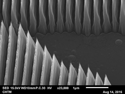
Scanning Electron Microscope (SEM) image of nanogratings magnified by 23,000
The Nuclear Magnetic Resonance (NMR) sensor uses a nanostructured diamond chip in a picoliter (pL) solution. The NMR detection sensitivity depends on the number of nitrogen-vacancy (NV) centers that are located close enough to the diamond surface to sense external spins.
To increase this number, the diamond surface is lithographically structured with dense, high-aspect-ratio nanogratings to enhance the sensor–analyte contact area by ≳15×. The nanostructure sidewalls are then doped with a high density of NV centers.
The result is tens of millions of NV centers that are located close enough to the diamond surface (5–20 nm) to detect the NMR spectrum from ~1 pL of fluid lying within the adjacent nanograting grooves. This leads to a corresponding boost in fluorescence signal and reduction in acquisition time.1
With further improvements in spectral resolution, this platform could enable a wide variety of applications in biochemistry, including pharmacodynamic studies of metabolites and natural products and high-throughput screening for drug discovery.
The sensor is structured to increase the total contact area with the sample. This allows for sensitive detection of sub-nanoliter volumes of trace samples under a wide variety of environmental conditions. In addition, this technique is capable of being employed in low magnetic fields to detect nuclear quadrupole resonance (NQR), making it possible to detect extremely small sub-nanoliter volumes of liquid, gas, and thin-film samples over a wide range of ambient temperatures without the need for large cryogenic magnets.2
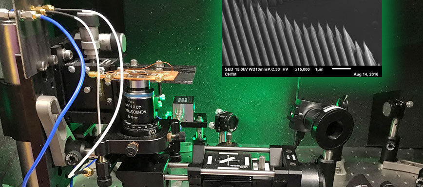
Nuclear Magnetic Resonance Spectroscopy setup, one of three at CHTM
Sources:
1. Nature Communications, doi:10.1038/s41467-017-00266-4.
2. STC.UNM Filing Abstract
Learn more about the research:
Nanoscale physics research team performs magnetic resonance spectroscopy on a diamond chip